When I started my PhD at McGill University, in 1973, I was thrilled to discover that the university had a library devoted just to ornithology: the Blacker-Wood Library of Zoology and Ornithology. This library was a primarily large room located in the basement of the university’s immense Redpath Library just down the road from the Biology Building. Besides a fabulous collection of contemporary books and journals about birds, the library also had a rare book room full of treasures like the feather book of Dionisio Minaggio. It also had a hand-coloured copy of John Ray’s Ornithology of Francis Willughby published in 1776 and ostensibly given as a present to Samuel Pepys, the famous 17th century diarist [1].
I remember the librarian, Eleanor MacLean, telling me that the library subscribed to more than 100 ornithological journals and newsletters. That seems like a lot but they did get the separate bird journals published by many of the provinces in Sweden, as well as a dozen or more in foreign languages that I could not read, so that total might not have been far off. It was a real treat for me to spend every Friday morning poring over the latest acquisitions. While I love the convenience of the internet, and rapid search engines, there was something oddly rewarding about opening the latest issues of journals from around the world, all devoted to the study of birds.
That library was founded in 1920 by Casey Wood, a Canadian-American [2] ophthalmologist who was also an avid—OK, obsessive—book collector. On Friday (20 April), I will be going to Montreal to participate in a symposium celebrating his contributions to the library in particular and to ornithology in general.
Wood wrote several papers about birds but his signal ornithological work was a little-known volume called The Fundus Oculi of Birds, published in 1917. Because Wood was an ophthalmologist and immensely curious, he thought it might be interesting to use the tools of his trade to study the eyes of birds. To do that, he employed the techniques that he had used in his practice, and further developed some procedures for studying the eyes of birds. His main interest was the structures of the fundus oculi, at the back of the eye—the retina, the fovea(s), the nerves and the pecten.
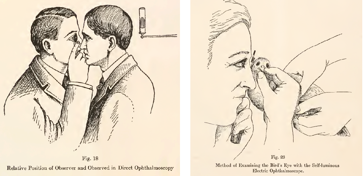
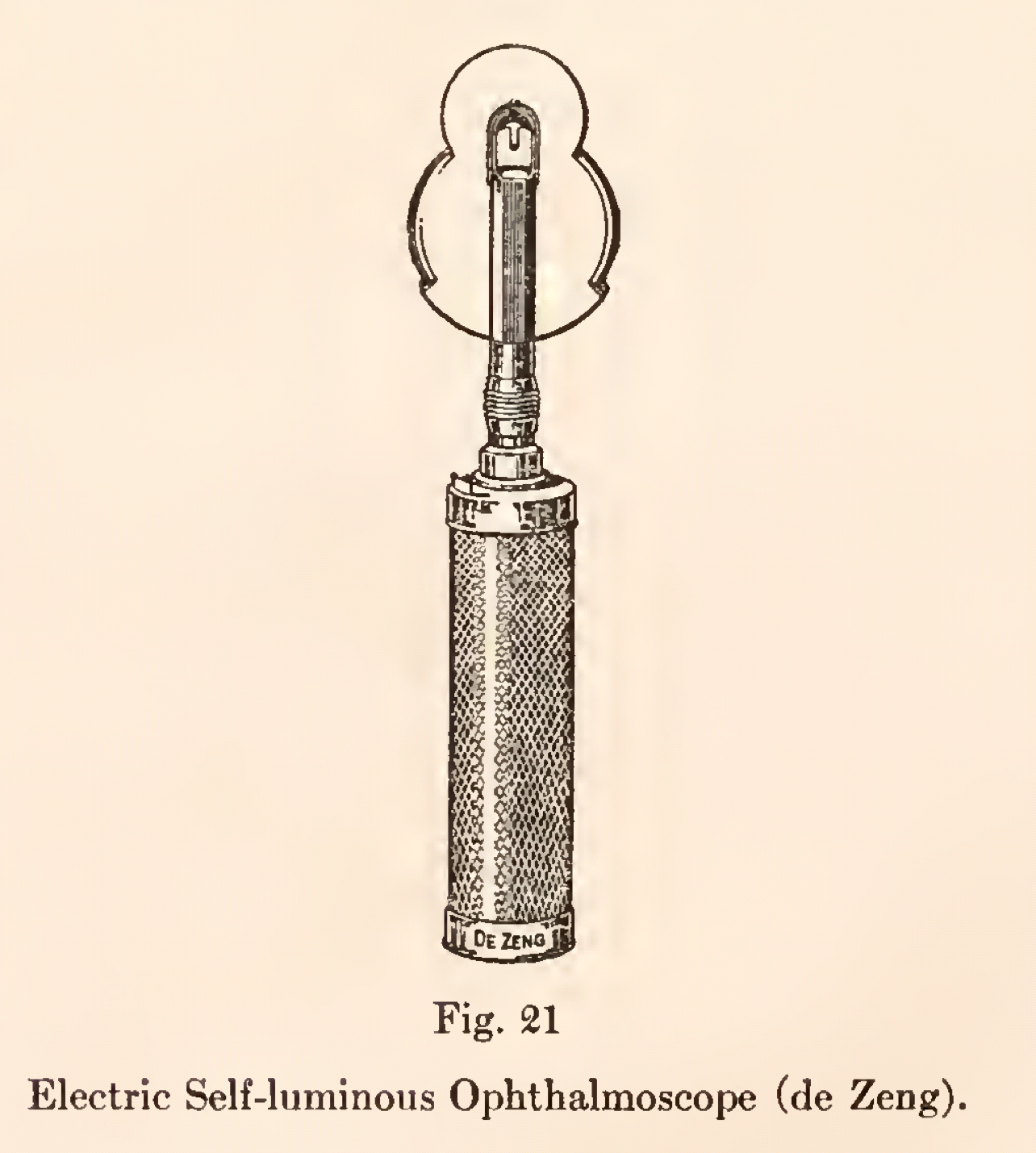 Wood realized early on that his observations would be most useful if he could somehow illustrate what he was seeing through his ophthalmoloscope. In those days, photography was simply not advanced enough to provide enough depth of focus:
In spite of recent advances in that direction, attempts to reproduce the colored (ophthalmoscopic) appearances of the fundus by photography have so far failed. [3]
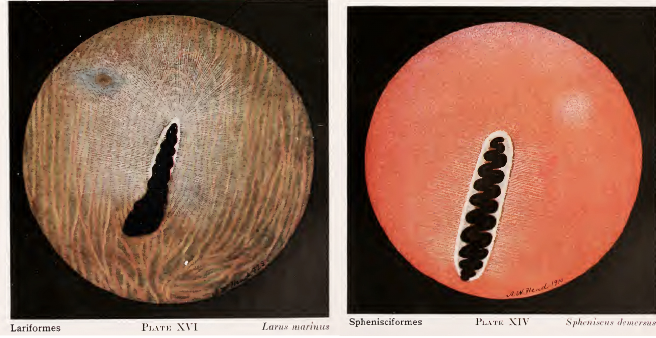
Instead he employed the talents of Arthur William Head [4], a celebrated UK artist, to draw and paint the fundus oculi of more than 80 bird species. Wood does not record how Head did this—did Wood train Heath to use the opthalmoscope or did he roughly sketch what he saw and then work with Head to make Head’s illustrations match what Wood saw? Wood says that it takes a lot of experience to be able to use the ophthalmoloscope but maybe he was able to teach Heath his techniques. Wood also says that his methods allowed only a short period of observation of the fundus oculi so it is remarkable how detailed these illustrations are, however they were produced.
Perhaps surprisingly, Wood took a very modern approach to his subject, as he attempted to examine at least one species in each avian order to see if there were patterns related to the bird’s way of life—an early example of what we now call comparative analysis.
The oculi of Birds exhibits a great variety of areas of distinct vision, and these correspond closely to the habits and habitat of these animals especially their methods of obtaining food, of escape from enemies, of migration, of reproduction, etc. [3]
But he also wanted to determine whether the fundus oculi might be a useful taxonomic tool, helping to define the evolutionary relationships among species. To that end, he sampled and illustrated the fundus oculi of birds from 24 of the Orders of birds that were recognized in those days, missing only 5 of the Orders that we (or at least some authorities) recognize today (Turniciformes, Craciformes, Gabuliformes, Coliiformes and Musophagiformes). There was a bit of a taxonomic signal there, in Wood’s opinion, but not clear enough to be useful.
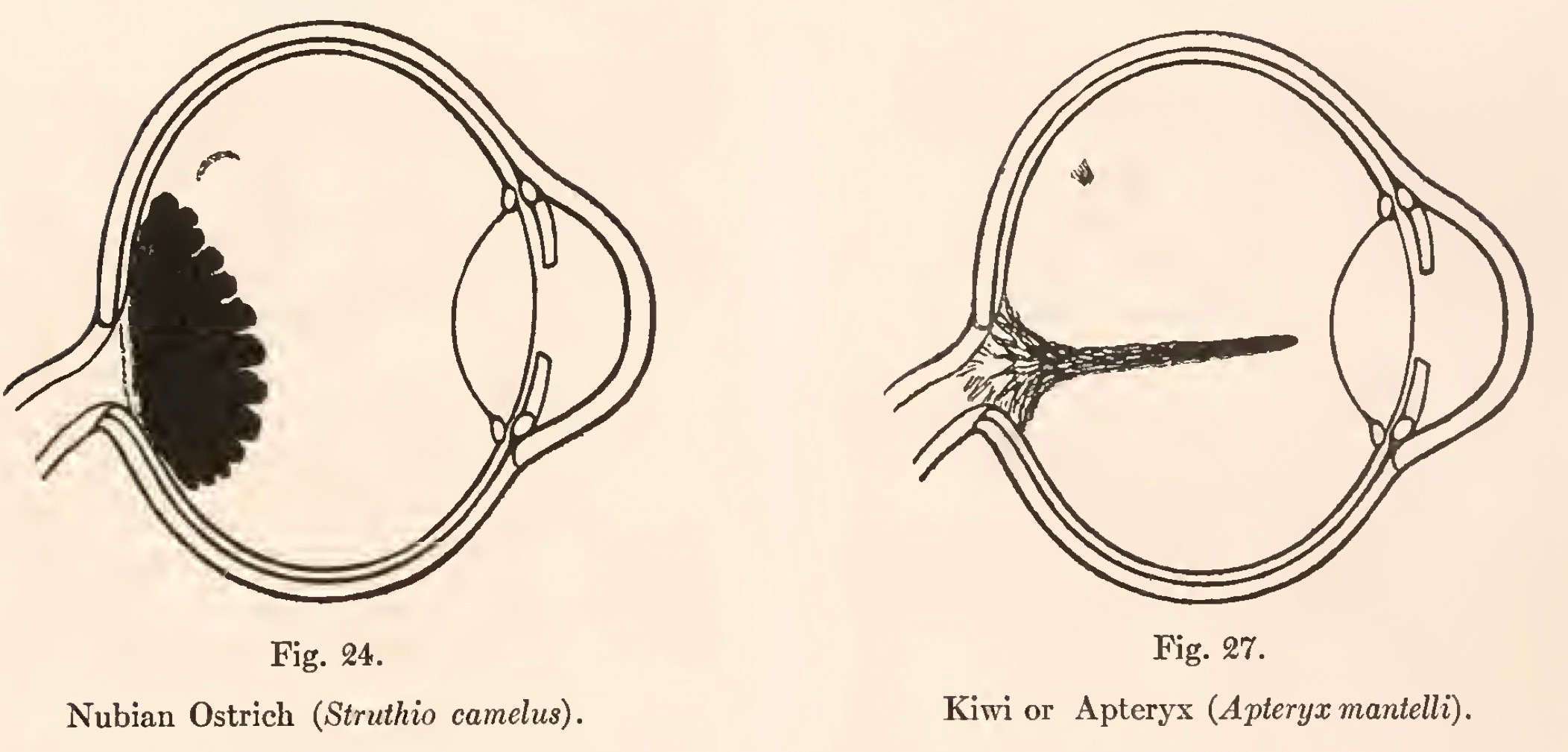
Instead, Wood showed nicely that the pecten size and shapevwere likely related to visual acuity, and that some birds have two foveas, related either to visual acuity or more likely the need to switch from binocular to monocular vision while foraging. Even today, a century later, we still do not have definitive studies of these traits in the fundus oculi, as far as I can tell.
Wood also found that the pecten degenerated in domesticated birds suggesting to me that it is a costly structure that becomes smaller under relaxed selection when visual acuity is no longer important for foraging or survival.
Reading Wood’s Fundus Oculi Book for my talk in Montreal reminded me once again of the tremendous value in reading the older literature once in a while. I have been interested in avian vision for about 25 years but had never really thought or read about the pecten before. To the best of my knowledge, many of the patterns that Wood described have not been further investigated even though we now have all of the tools needed to measure and quantify the structures he described and illustrated. Wood’s book is not an easy read as it is largely descriptive and lacks focus (no pun intended). There are enough gems buried there, though, that anyone interested in the vision of birds might want to have a look at this interesting book.
SOURCES
- Montgomerie R, Birkhead TR (2009) Samuel Pepys’s hand-coloured copy of John Ray’s ‘The Ornithology of Francis Willughby’ (1678). Journal of Ornithology 150:883-891 DOI 10.1007/s10336-009-0413-3
- Wood CA (1917) The Fundus OCuli of BIrds ESefially as Viewed by the Ophthalmoloscope: A Study in Comparative Anatomy and Physiology. Chicago: Lakeside Press. Available here
Footnotes
1. Pepys’s copy of Willughby and Ray’s Ornithology: see Montgomerie and Birkhead (2009)
2. Casey Albert Wood: (1856-1942) was born in Wellington, Ontario, attended high school in Ottawa, and got his medical degrees from McGill University. He eventually settled in Chicago where he practiced ophthalmology. See here for details
3. Quotations: from Wood 1917 page 7
4. Arthur William Head: (1861-1930) was a celebrated artist who lived in London, England, see here for examples of his work
Nice post! Libraries and librarians matter and the gems from the past are all too often forgotten! Wonderful to see you last week at AOS Tucson!
Taylor accepted the award in April at the American Ornithological Society ’s annual meeting in Tucson, Arizona, where he gave a plenary talk titled “A bird’s eye view of the origin and maintenance of biodiversity.”